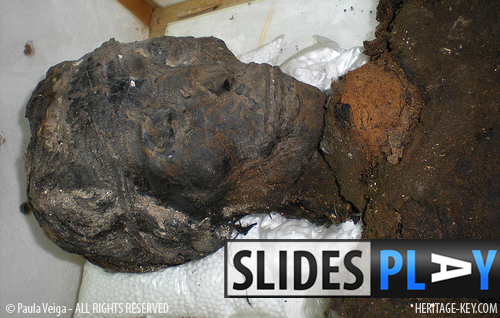
 We are just learning fresh news about research on King Tut’s mummy, in advance of tomorrow’s publication in the American Medical Journal of the results of the most recent DNA and other tests. Over the years, there have been many different theories, but now we can scientifically prove what killed the Boy King, his parentage, and other health conditions affecting him at the time of his death.
Early Research
KV62 – Tut’s tomb – was discovered by Howard Carter in 1922. Multiple attempts at proving kinship between various royal mummies have been made since then, including tests by Connolly (1976), Flaherty (1984) and Harrison (1969). In the case of Tutankhamun and Smenkhare, these tests have included estimates of both mummies’ blood groups in order to compare them.
Both mummies share the same rare blood type (group A2, and both with the serum antigen MN), suggesting close consanguinity.
In 2000, Tutankhamun was due for testing again. This time, a Japanese team would attempt tot extract DNAfrom the mummy. Shortly after the announcement was made, the Egyptian government decided to revise the granted permissions, and the planned geneaology and paleopathology research was cancelled.
The Two Fetuses – King Tut’s Daughters
In the case of the two fetuses found in KV62, the DNAtest confirmed a theory. The two girls have different body shapes, but their DNAwould quickly prove if they really are sisters, and even twins – as suggested by Connolly. He believes the difference are symptoms of a rare event in which one twin consumes more nutrients from the mother than the other, and is therefore born much bigger and stronger. Dr. Connolly explained this theory to me himself, when I was attending Manchesters KNH Centre for Biomedical Egyptology in 2008.
Premature or severely ill newborn babies hardly ever survived in Ancient Egypt, and often a child died in the mother’s womb. It is very probable that Tutankhamun’s daughters are an example of this, as they were far from full-grown: they died at five and six months gestation.

The First CT-scans
In 2005, Madeeha Katthab, Dean of the Medical Department of Cairo University, together with his team and aided by specialists from Italy and Switzerland, performed a CAT scan of Tutankhamun’s mummy (using equipment kindly donated by National Geographic and Siemens Medical Solutions).
Based on these scans, in 2007, Dr. Benson Harer was the first to suggest that King Tut’s early death might have been linked to the pharoah’s leg.
An interesting detail worth mentioning from Harer’s research is that if Tutankhamun had a deficient immune system, ancient Egyptians were already knowledgeable in this area. Black cumin oil was known in Egypt as a stimulant and as an reinforcing agent for the immune system. When the tomb of Tutankhamun was first opened, archaeologists discovered a bottle of black cumin oil, no doubt gifted to the King to ensure a painless afterlife.
Where Was His Willy?
Not related, but too funny not to mention, is that Tutankhamuns lost phallus had been hiding in the sandbox (the sand around the mummy) since the 1960s. The missing member generated a lot of controversy; it is clearly present in Burton’s photographs, but at a certain point disappears from the (not Burton) picture. King Tut’s member was rediscovered by Dr Hawass in 2006, who found that it had never left the sandbox after all.
The Murder Conspiracy
Speculations about Tut’s cause of death – and his missing penis – started in 1968, when a team from Liverpool University, led by Professor Ronald Harrison, X-rayed Tutankhamun’s body in his tomb. These images revealed a possible blunt force injury to the back of the King’s head and the presence of what looked like bone fragments inside the skull.
I learned from Dr. Connolly (I also had a glance at the original X-rays myself) that the bone fragments inside Tutankhamuns skull, commonly called the vault, were small fragments from the smallest bones we have in the skull next to the eyes and nose (nasal, lachrymal and palatine). These tiny bones break easily, so could have well been damaged in the process of mummification.

Break a Leg
The pathological condition that King Tut suffered in his leg was a bone inflammation that, according to the recent released article, was enhanced by his weakened immunity system.
Sir Marc Armand Ruffer studied several Egyptian bodies. Writing in 1921, he described the typical condition of leg bones: “In contrast to the spine, the femurs showed, as a rule, but slight lesions, and even these did not occur often. Altogether only nine femurs showed any lesions, the most pronounced of which, at the upper end.”
Osteomyelitis is the inflammation of the marrow cavity; thirty-one cases were noted on twenty-six individuals from the Predynastic cemetery at Naga ed-Der. The tibia and maxilla are the most frequent affected bones, with ten examples known for the tibia.
Elliot Smith and Wood Jones nevertheless concluded that inflammatory diseases of bone were rarely seen in ancient Egyptian skeletons. New data published show they also found that “the left second metatarsal head was strongly deformed and displayed a distinctly altered structure, with areas of increased and decreased bone density indicating bone necrosis.”
One Pharoah, 130 Walking Sticks
The deformities found in King Tut’s foot indicate that the disease was ongoing at the time of his death. Since Howard Carter discovered 130 whole and partial examples of sticks and canes in the kings tomb, we might say that ancient Egyptians prepared themselves well for the afterlife. These finds support the hypothesis of a walking aid being a necessity for the young kings travel after death. Some of the canes found in KV62 are worn, which consolidates the idea that he must have needed some kind of cane to walk.
A Rather Rare Physiognomy?
According to Dr. Corthals, genotype defines phenotype, so, the application of DNA testing can also help to determinate if the strange physiognomy observed in depictions of Akhenaten and his children – possibly including Tutankhamun – may derive from a genetic corridor set up by his ancestors, meaning a genetically-inherited feature.
During the New Kingdom, the environment did not changed substantially enough to disrupt a genetic trace, so it would be possible to confirm Akhenatens genetic characteristic and its passage to his offspring.
The newly published article in JAMA states that “a Marfan diagnosis cannot be supported in these mummies.” This means that all the theories suggesting feminine traits in this dynasty crumble, as science – once again – proves them wrong.
The full text reads: “Macroscopic and radiological inspection of the mummies did not show specific signs of gynecomastia, craniosynostoses, Antley-Bixler syndrome or deficiency in cytochrome P450 oxidoreductase, Marfan syndrome, or related disorders.”
Another fascinating part of this study is that, in many cases, DNA analysis (see how they take the samples in this photo preview of ‘King Tut Unwrapped’) provides information that makes it possible to detect an otherwise invisible infection.
Akhenaten is the Father of King Tut (Probably)
The research also concludes that the KV55 mummy, who is most probably Akhenaten, is probably the father of Tutankhamun. According to the latest scientific data published today in the JAMA (Ancestry and Pathology in King Tutankhamun’s Family), “Syngeneic Y-chromosomal DNA in the Amenhotep III, KV55, and Tutankhamun mummies indicates that they share the same paternal lineage.”
KV55 is thought to have housed Akhenatens body and the article clearly states that “…the KV55 mummy, who is most probably Akhenaten, father of Tutankhamun. The latter kinship is supported in that several unique anthropological features are shared by the 2 mummies and that the blood group of both individuals is identical.”
However, this does not mean the identification of the mummy in KV55 is conclusive.
A Mean Case of Malaria
Last but not least, after PCR amplification of DNA samples they found indicative proof of at least a double infection with the P falciparum parasite (malaria) in Tutankhamun, as well as the mummies of his ancestors Thuya and Yuya. The type diagnosed is malaria tropica, the most severe form of this disease. Although Tut’s relatives suffered from malaria as well, they lived much longer than him. Apossible explanation is that, although they all lived in a malaria endemic area, the ladies did not suffer from the same other pathologies (almost all of the above) that Tutankhamun did.
The photos of Tut and his daughters I’ve mentioned can be consulted in a publication by Leek F. The Human Remains from the Tomb of Tutankhamun. Oxford, UK: Tutankhamun Tomb Series V; 1972. More bibliography on the serological tests, previous tests done on Tut and his daughters can be browsed in the JAMA article references list.


